



Breast Cancer Screening
There are different imaging methods adopted as breast cancer screening tools, including mammography, breast ultrasound and breast magnetic resonance imaging (MRI). Each modality has its own advantages and limitations. All women who consider breast cancer screening should discuss with her doctor to make an informed decision.
Mammography
- Mammography is a common screening tool for breast cancer and it is an X-ray examination of the breasts. Studies have shown that mammography is effective in detecting early-stage breast tumours that cannot be easily seen or felt.
- During the procedure, women will expose her upper body. By gently compressing the breasts with the X-ray machine's flat plastic paddle, X-ray images of the breasts can be taken at different angles.
- Women may feel discomfort or even pain during breast compression, but that part of the mammography usually lasts only a few seconds, and is essential to improve image quality and diagnostic accuracy.
- Most countries adopt 2D mammography as the standard for breast cancer screening. Currently, there is insufficient evidence to support using 3D mammography as a population screening tool to reduce breast cancer mortality. However, women with dense breast tissue might benefit from 3D mammography, please discuss with your doctor on screening modalities most suitable for you.

Breast ultrasound
- Breast ultrasound is a test that visualises the internal environment of the breast by transmitting high frequency sound waves to the breast tissue and converting the reflected echo into image.
- Breast ultrasound is used as an adjunct to mammography in women with radiologically dense breasts, to depict small breast cancers not visible on mammography.
- Women may be advised by the doctor to receive additional breast ultrasound if clinically indicated.

Breast magnetic resonance imaging (MRI)
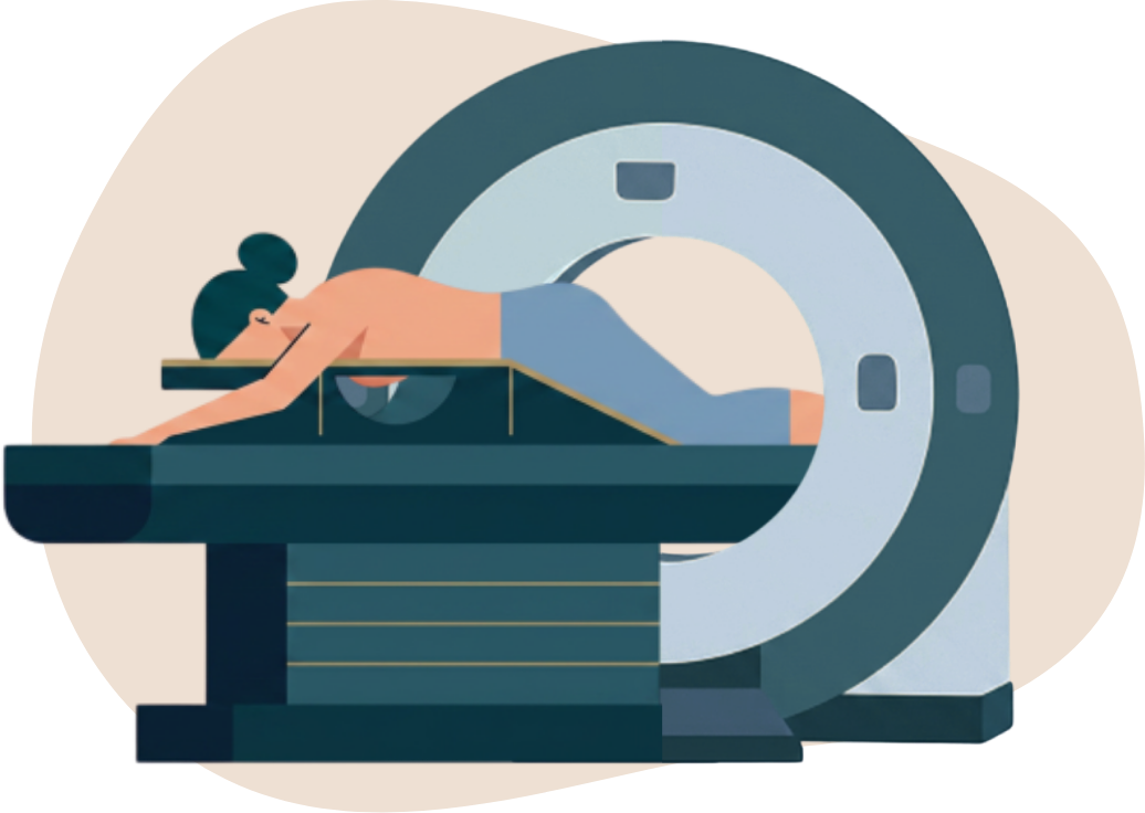
- Breast MRI is a test that produces detailed images of the breast internal environment by using strong magnetic field, radio waves and computer technology.
- Women with confirmed BRCA1/2 deleterious mutations or who had radiation therapy to the chest for treatment between age 10 and 30 years may be advised by the doctor to consider breast MRI, as an adjunct to mammography for higher sensitivity.
Points to note
- All screening tests have their limitations and they are not 100% accurate. They bear potential risks, e.g. false-positives, false-negatives, over-diagnosis and unnecessary treatment.
- All women who consider breast cancer screening should discuss with doctors on the potential benefits and harms before undergoing screening to make an informed decision.
- Under the Government-subsidised Breast Cancer Screening Pilot Programme Phase II, the provision of 2D mammography and/or ultrasound, if indicated, will be subsidised. However, you should also be aware that for any additional services to which participants may be referred, such as magnetic resonance imaging (MRI) screening of the breast, genetic testing / counselling, and consultation with specialists (e.g. breast surgeons, gynaecologists) on breast cancer risk reduction strategies, clients would pay out of pocket. You are advised to consult your doctor on the potential costs of these services.